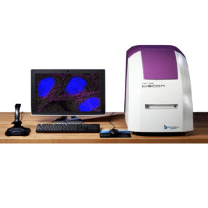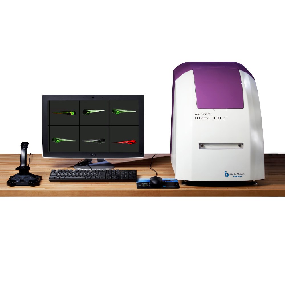
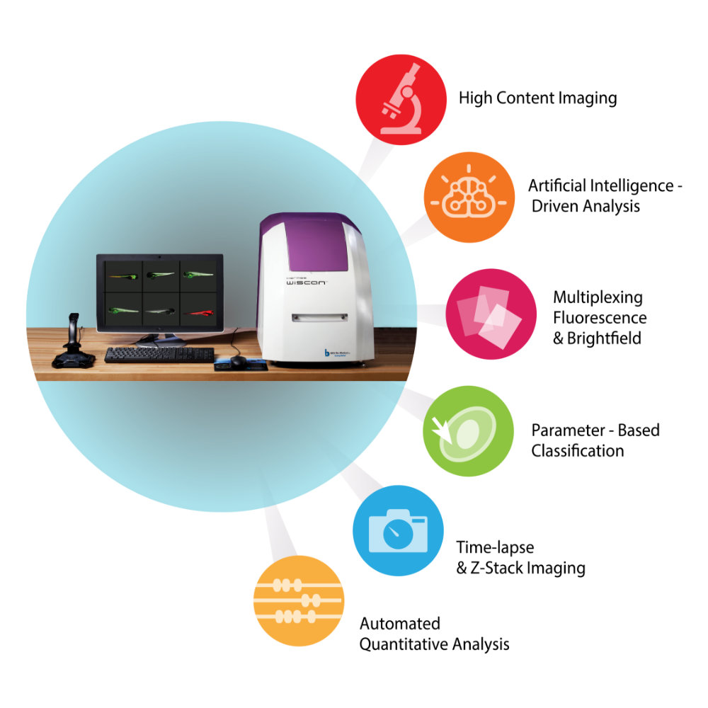
WiScan® Hermes High Content Imaging System
- Phenotypic Screening
- Spheroids and 3D Models
- Rare-event detection
- Cytometry
- Cell Counting
- Protein Expression
- Cell Morphology changes
- Cell cycle tracking
Watch the webinar here
Easily generate publication-quality images at high throughput speeds
WiScan® Hermes is a cost-effective system that is both sophisticated and flexible, offering up to 7 fluorescence colours, bright field option, and a large range of air objectives. The system can accommodate a variety of multi-well plates and sample formats (slides, dishes…) and offers environmental control for live cell assays.
Versatile
Whether you are performing assay development, compound screens, transfection assays or looking at a few samples in great detail, and whether you are using 3D models (such as spheroids, Organoids), Zebrafish imaging, primary cells, fixed cells or live-cell imaging, WiScan® Hermes is the solution for you.
Reliable
WiScan Hermes, IDEA Bio-Medical’s automated imaging system for high content screening (HCS) provides the unique combination of the two contradicting primary functions of automated microscopy: Image quality and acquisition speed.
Robust
WiScan® Hermes’ mechanisms are based on patents, creatively designed to meet heavy-duty operation demands (24/7) with full process robustness.
Flexible
WiScan® Hermes is a cost-effective system that is both sophisticated and flexible, offering 7 fluorescence colours, bright field option, and a large range of air objectives. The system can accommodate a variety of multi-well plates and sample formats (slides, dishes…) and offers environmental control for live cell assays.
Intuitive
It doesn’t matter if you are a beginner or an experienced microscopist, Hermes easily allows you to look deeper into your samples. The system is intuitively operated. Its built-in applications are extremely easy to use and are operated at the push-of-a-button.
| Features | Content/Description |
| 3D reader | EPI-fluorescence inverted optics mounted on XYZ (patented) linear scanner |
| Auto Focus | Patented ultra-fast laser-based Auto Focus with 100nm resolution |
| XY motion | Accurate positioning with 200nm repeatability |
| Illumination sources | 7 optional LED sources (DAPI,CFP,GFP,YFP,RFP,mCherry,CY5). Transmission: White LED source |
| Optical Filters | 2 emission filters and compatible dichroic filters (automatically exchanged) |
| Objectives (Air) | Choice of air objectives in the range: 2X to 60X, high NA. |
| Camera | High sensitivity CCD camera with 1.3MPixel resolution |
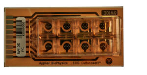
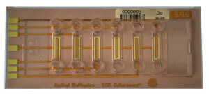
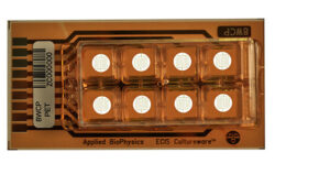
| Sample format | Supports full-area screening of 6-1536 well plates Supports slides, microarrays, 35 mm dish formats. U-shaped bottom plats are optional. |
| Computer | PC with Windows® operating system and touch screen. |
| Enclosure | Allows operation in fully lit areas |
| Desktop standalone platform | 47 W x 72 D x 57 H (cm), 18.5 W x 28.5 D x 22.4 H (inches) With plate cover closed |
| Certification | CE, UL |
Live Cell Imaging Module:
| Features | Content/Description |
| Live cell imaging | Allow long time lapse experiments. Sample does not move during most of the scanning process. |
| Live cell conditions |
|
Object Mapping (Rare Event Detection) Module:
| Features | Content/Description |
| Object mapping for rare events detection |
|
| Analysis tools | WiSoft® based image processing tools for object definition and detection in the first low magnification scan and further tools to analyze the detected objects with high magnification |
| Objectives | Automatic objective exchanger of up to 3 objectives at a time |
| Throughput | Enhances the throughput of high magnification scanning of rare objects |
| Statistical tools | Provides automatic decision resulting in efficient screening for minimum required objects in a well |
High Throughput (enhanced acquisition speed) Module:
| Features | Content/Description |
| Combination of HCS and HTS | Ultra-fast High Content Screening unit |
| Image acquisition speed | >10 images per second (depends on experiment conditions) |
| Throughput | A full 96-well plate screening using 10X magnification with a single field per well, three color fluorescence, 150 ms total exposure time per field, runs in ~2 minutes. With some applications, this will include data processing and analysis results simultaneously with image acquisition. |
Automation Module (interfacing to external robotic arms):
| Features | Content/Description |
| Remote accessibility | Protocols ( applications and screening parameters ) execution and management |
| External loader compatibility (Robot/manipulator) |
|
| External loader(Robot/manipulator) integration | Full integration support |