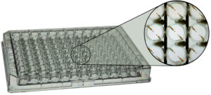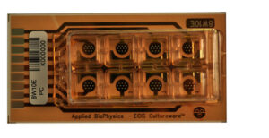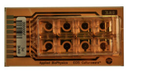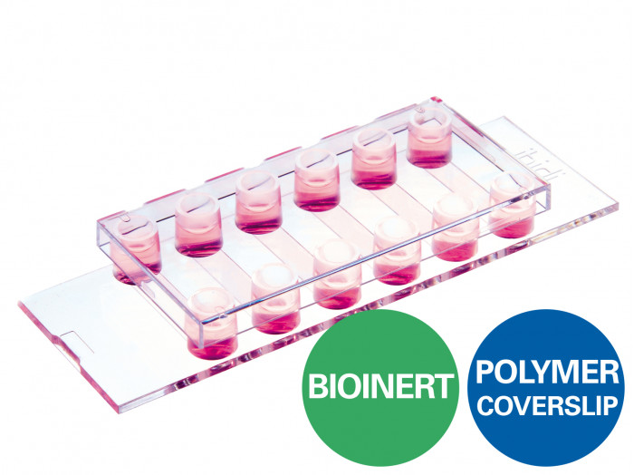
µ-Slide VI 0.4 Bioinert

A 6 channel μ-Slide with a non-adherent surface for flow experiments and fluorescence microscopy
Applications:
- Real-time imaging under either static or flow conditions
- Generation and long-term culture of 3D cell aggregates (e.g., spheroids and organoids) without any scaffold
- High resolution fluorescence microscopy of organoids, spheroids, embryoid bodies (EBs), and cells in suspension
- Background-free analysis of cell-cell interactions
- Prevention of stem cell differentiation due to attachment
- Culture of suspension cells in a permanently unattached state
- Self-assembly tumor spheroid formation assays
- 3D tumor spheroid models
Technical Features:
- Ready-to-use without prior surface treatment or preparation
- No adsorption, coating, or binding of proteins, antibodies, enzymes, and other biomolecules
- Non-cytotoxic, biologically inert, and non-degradable
Specifications
Bioinert Surface
| Bioinert surface thickness | 200 nm |
| Bioinert surface material | Polyol-based hydrogel |
| Protein coatings | Not possible |
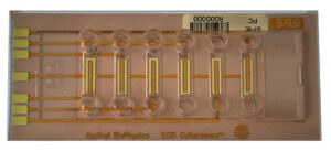
µ-Slide VI 0.4
| Outer dimensions (w x l) | 75.5 x 25.5 mm² |
| Adapters | Female Luer |
| Number of channels | 6 |
| Channel volume | 30 µl |
| Channel height | 0.4 mm |
| Channel length | 17 mm |
| Channel width | 3.8 mm |
| Volume per reservoir | 60 µl |
| Growth area | 0.6 cm² per channel |
| Coating area using 30 µl | 1.2 cm² per channel |
| Bottom: ibidi Polymer Coverslip with Bioinert surface | |
Specifications
Bioinert Surface
| Bioinert surface thickness | 200 nm |
| Bioinert surface material | Polyol-based hydrogel |
| Protein coatings | Not possible |
µ-Slide VI 0.4
| Outer dimensions (w x l) | 75.5 x 25.5 mm² |
| Adapters | Female Luer |
| Number of channels | 6 |
| Channel volume | 30 µl |
| Channel height | 0.4 mm |
| Channel length | 17 mm |
| Channel width | 3.8 mm |
| Volume per reservoir | 60 µl |
| Growth area | 0.6 cm² per channel |
| Coating area using 30 µl | 1.2 cm² per channel |
| Bottom: ibidi Polymer Coverslip with Bioinert surface | |
Variant
Pack/Size
Price
Qty
µ-Slide VI 0.4 Bioinert: #1.5 polymer coverslip, surface passivation with Bioinert, Sterilized
15 / Pack
£683.80

µ-Slide VI 0.4 Bioinert, Bulk Box 120: #1.5 polymer coverslip, surface passivation with Bioinert, sterilized, 8 per tray, 15 trays
120 / Pack
£4,440.59
µ-Slide VI 0.4 Bioinert, Bulk Box 90: #1.5 polymer coverslip, surface passivation with Bioinert, sterilized, individually packed
90 / Pack
£3,538.58